



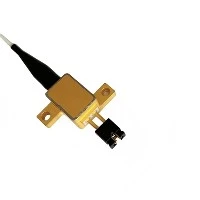
Intrafocal treatment using a 1470 nm semiconductor laser and bare fiberHypertrophic scar and keloid is a new and effective minimally invasive technique.
Hyperplastic scars and keloids are caused by abnormal wound healing and respond differently to many available treatments. In recent years, the development of laser therapy has shown a wide range of clinical applications, including scar treatment. We studied the efficacy of 1470 semiconductor laser combined with fiber optics in the intraluminal treatment of hypertrophic scar and keloid
To evaluate its safety and effectiveness as a new minimally invasive treatment in controlling scars and reducing scar volume. We conducted a prospective cohort study that included 21 patients with hypertrophic scars (HS) (n=9) and keloids (n=12) due to various etiologies. Patients receive one to three courses of treatment.
Heal. Vancouver Scar Scale, Doppler ultrasound, skin elasticity tester, Mexameter, and PeriCam-PSI were used for comprehensive evaluation. The mean reduction in scar thickness was 0.308± 0.138 cm (p< 0.001). In particular, these two groups showed a significant reduction in the scar thickness of HS and keloid by 27.7% and 28.2%, respectively. Scar hardness in the HS group was significantly improved by 1.2% (p< 0.05), keloid group improved by 0.4% (p=0.26). Keloid pigmentation was significantly reduced by 21.3%. Blood perfusion was reduced by 29.6% in the HS group and 22.7% in the keloid group. The total VSS score of the HS group was improved by 42%, and the total VSS score of the keloid group was improved by 37.9%. There were no adverse reactions such as hyperpigmentation or hypopigmentation, skin infection, or recurrence. This study showed that, based on both subjective and objective analysis, the intrafocal 1470nm semiconductor laser and bare fiber significantly improved hypertrophic and keloid scars, and demonstrated that this type of laser therapy is a safe and effective minimally invasive treatment option.
Hypertrophic scars (HS) and keloids (K) are the result of abnormal wound healing and scarring. Pathological scars are characterized by persistent inflammation and histological features such as fibroblast proliferation, neovascularization, and collagen deposition. The clinical feature of HSK is an overgrowth of tissue at the edge of the original wound. HS usually occurs within a few months when the scar is in tension, including the sternal area, around the joint, and can persist along the edges of the original wound. Keloid scars are more common in individuals with darker skin and can form within a few years after minor trauma, as well as in areas such as the earlobes, shoulders, and chest. Keloids are often symptomatic, associated with pain and itching, and can grow well beyond the original boundaries of the wound. HSK occurs through various injury mechanisms, including skin lesions, burns, surgery, injections, and dermatitis. Among them, deep burns are reported to be the main cause of HSK.
First, non-surgical treatment is preferred. However, if that fails, surgery, laser, and radiation therapy are considered favorable treatment options. 10 Although minimally invasive surgery has good results for softer, thinner scars, it is difficult for harder and thicker scars. Local drugs are difficult to penetrate, injectable therapeutics are blocked by fibrotic tissue, and multiple treatments are required, often with limited effectiveness. Traditional laser diffusion also has limited effectiveness in thicker scars.
HSK responds positively to chemotherapy drugs, but side effects include pain, ulcers, burning, and pigmentation. Surgical removal of HSK is still an option, but unfortunately, the long-term outcome can be poor and the risk of scarring recurrence is high. Currently, no one method shows an overall advantage and recent publications have shown conflicting results. The treatment of HSK remains a clinical challenge. 11125,6,10 This study builds on our previous positive experience using a 1470 nm semiconductor laser and fiber optic system to treat red, swollen, and infected keloid cells.
We demonstrate that a 1470nm semiconductor laser can generate local heating effects over a narrow range through a fiber optic system. Fibrotic scar tissue evaporates rapidly, causing fine
Cytolysis, necrosis, and coagulation lead to tissue ablation and control inflammation. In addition, the 1470nm laser is able to coagulate blood vessels, reducing blood supply and local tissue blood vessel formation. 1314-16 The objective of this study was to evaluate the efficacy and safety of the 1470nm laser and fiber optic system, a novel scar volumetric reduction therapy, and to evaluate its efficacy in the treatment and improvement of HSK.
The study employed a graded non-ablation laser lasemar1500 (EUFOTON, Trieste, Italy) whose semiconductor gallium arsenide (GaAs) emits a laser at a wavelength of 1470nm. The energy is transmitted through disposable optical fibers with a diameter of 300 microns. Set the power output according to the scar hardness, penetrate the scar with the least resistance, generally starting from 3w, and gradually increasing to the maximum power output of 6w when necessary, with the maximum injection of 999.9KJ/cm2. After the laser device is started, the fiber transmits energy and penetrates the scar for 1 to 4 channels. The puncture points surround the scar, with each point separated by 2-4 mm. This is repeated every one to two months until the results are satisfactory to the clinician and patient.
Contact information:
If you have any ideas, feel free to talk to us. No matter where our customers are and what our requirements are, we will follow our goal to provide our customers with high quality, low prices, and the best service.
 Email:info@loshield.com
 Tel:0086-18092277517
 Fax: 86-29-81323155
 Wechat:0086-18092277517


